Break Down the Damage. Reboot the Healing. Avoid Tendon Surgery.
Stuck in a healing dead-end? If physical therapy has stalled and steroid shots have worn off, your tendon is trapped by dead scar tissue or hardened calcium. Our ultrasound-guided Percutaneous Needle Tenotomy (PNT) breaks up the damage from the inside out, triggering a massive healing response.
Message Us to Book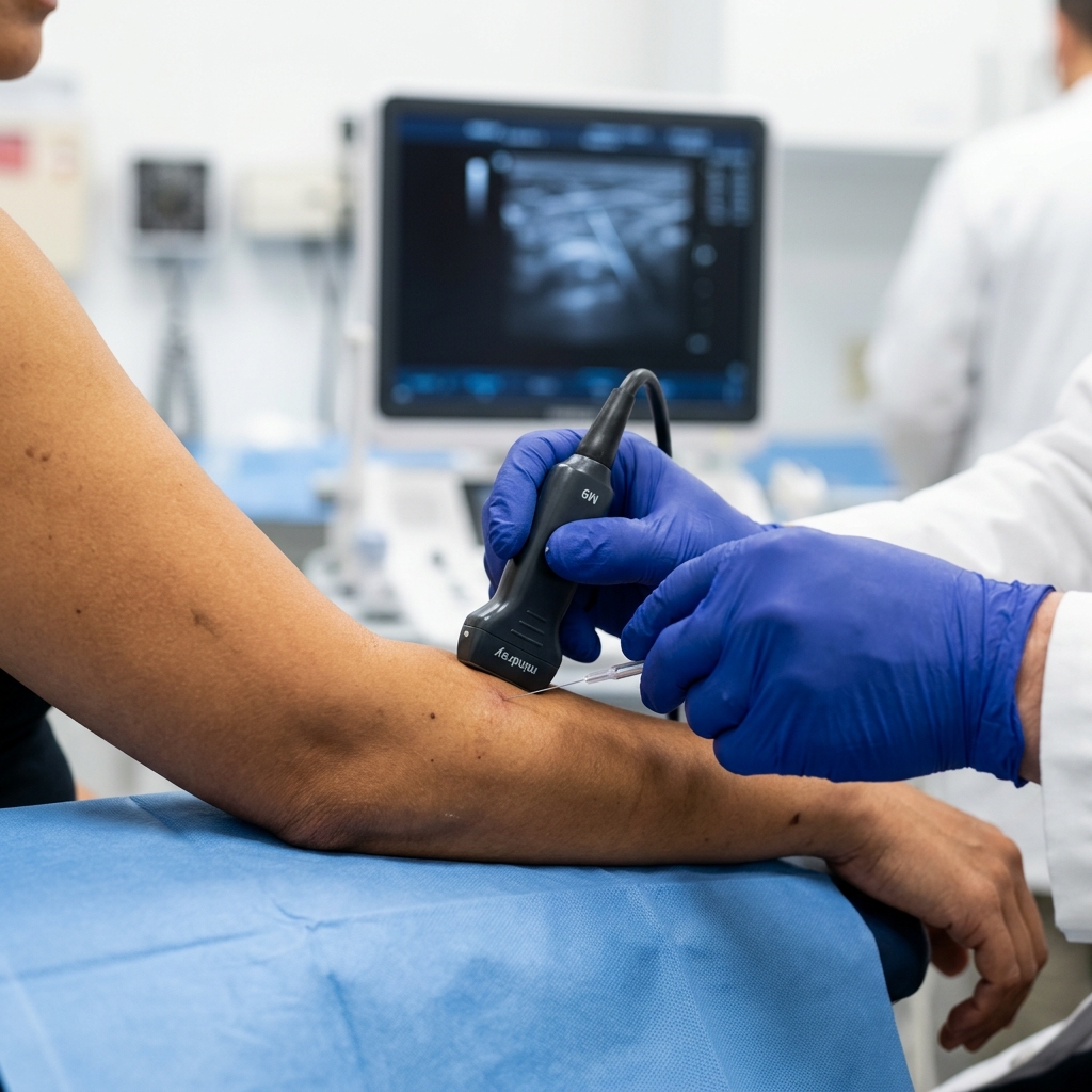
Escaping the Healing Dead-End
When you have suffered from Tennis Elbow, Achilles pain, or Plantar Fasciitis for more than six months, the problem is no longer simple "inflammation." Your body tried to heal the injury but failed. The healthy, flexible tendon tissue has been replaced by a dense, disorganized mass of dead scar tissue. In the shoulder, this tissue can literally turn into hardened, chalk-like calcium. At this stage, your tissue is acting like solid concrete. This is exactly why standard physical therapy stretches aren't working, and why steroid shots only hide the pain temporarily. You cannot stretch concrete, and steroids cannot resurrect dead tissue.
Before a surgeon cuts your joint open to scrape out the bad tissue, there is a highly advanced, minimally invasive alternative. Percutaneous Needle Tenotomy (PNT) acts like a micro-jackhammer. Under live ultrasound guidance, Dr. Rabara uses a fine needle to poke tiny, precise holes into the dead scar tissue, breaking it apart. This immediately signals your brain that there is a "new" injury, rushing fresh blood, oxygen, and growth factors to a zone that had zero blood flow. We literally "reboot" your body's natural healing process.
Chronic Tendon Conditions We Treat
Is Needle Tenotomy Right For You?
The Ultimate Alternative to the Scalpel
We reserve this procedure for patients who are incredibly frustrated. If you feel like your body has betrayed you by refusing to heal, this is your intervention.
You are an EXCELLENT candidate if:
- Your tendon pain (elbow, heel, shoulder) has lasted for more than 4 to 6 months.
- You have diligently tried physical therapy, but your progress has plateaued.
- Previously received a cortisone shot that felt great for 3 weeks, but the pain came roaring back.
- You have been told you need surgery, but cannot afford months in a cast or sling.
You may NOT be a candidate if:
- You just injured the tendon last week (requires basic rest and standard physical therapy first).
- Your tendon is completely and entirely torn in half (full-thickness rupture often requires surgical reattachment).
The Realities of Rebooting Your Tendon
Percutaneous Needle Tenotomy is incredibly effective, but it is not a "magic painkiller." We are actively initiating a healing response, which means things must get slightly worse before they get permanently better.
- The "Flare-Up" is Mandatory: We are intentionally causing micro-trauma to reboot the blood flow. For the first 3 to 7 days after the procedure, your tendon will feel highly sore, stiff, and inflamed.
- No Anti-Inflammatory Pills: Because we want the inflammation to heal the dead tissue, you are strictly prohibited from taking Ibuprofen, Celecoxib, or other NSAIDs for two weeks after the procedure.
- Biological Healing Takes Time: Unlike a steroid shot that hides pain instantly, tenotomy relies on your body growing new tissue. Noticeable relief typically becomes noticeable around 4 to 6 weeks.
The "Band-Aid" Procedure vs. Open Surgery
A surgeon cuts the skin open, slices into the tendon, and uses instruments to scrape out the dead tissue or calcium. Requires anesthesia, stitches, and high infection risk.
Dr. Rabara uses a tiny needle under ultrasound to break up the scar tissue and wash away the calcium through a pinhole in the skin. Zero stitches, minimal downtime.
A strong anti-inflammatory injected near the tendon to stop pain. In the Achilles, this actually weakens the tissue and increases snapping risk.
The Timeline to a Healed Tendon
The "Reboot" Phase
Your tendon will feel highly sore and stiff. This is fresh blood rushing to the dead zone. Rest the joint and avoid all heavy lifting.
Biological Rebuilding
Fibroblasts are laying down new, healthy tendon tissue. We introduce specific, gentle stretching and physical therapy protocols.
Return to Sport/Work
New tendon tissue has fully matured. Chronic pain has vanished. Confidently return to heavy lifting, tennis, gold, or running.
Clinical Science & Technical Details
For our medical colleagues and highly analytical patients, we provide these transparent technical details on the pathophysiology and interventional protocols.
Explore the Clinical Science: Tendinosis vs. Tendinitis and Angiofibroblastic Dysplasia
Chronic tendon pain (beyond 3-6 months) reveal an absence of inflammatory cells. Instead, the tissue exhibits 'Angiofibroblastic Dysplasia'—a degenerative process characterized by fragmented collagen, mucoid ground substance, and dysfunctional micro-vessels (neovascularization).
Percutaneous Needle Tenotomy (tendon fenestration) solves this structurally. The repetitive mechanical disruption of the fibrotic matrix using a needle incites controlled micro-trauma. This releases endogenous PDGF and TGF-β, converting a chronic, stalled degenerative lesion back into an acute, biologically active healing state.
Detailed Diagnostic Pathways: Barbotage for Hydroxyapatite Calcium
In Calcific Tendinitis, hydroxyapatite calcium crystals are deposited directly within the supraspinatus or infraspinatus tendons. The 'resorptive phase' is intensely painful, often requiring emergency intervention.
Dr. Rabara treats this utilizing Ultrasound-Guided Barbotage (Lavage). Using real-time sonography, we visualize the hyper-echoic calcium deposit and introduce a needle to break up the chalky deposit and aspirate fragments. This provides immediate decompression of the intratendinous pressure, offering rapid pain relief.
Advanced Systemic Screening and Sonographic Tendon Evaluation
Prior to tenotomy, Dr. Rabara performs an exhaustive Diagnostic MSK Ultrasound evaluation. We analyze the fibrillar echotexture to locate focal tendinosis or intra-substance interstitial tearing.
By visualizing the tissue dynamically, Dr. Rabara ensures we only target the disorganized scar tissue while meticulously preserving the surrounding healthy collagen fibers, guaranteeing absolute mechanical safety during the fenestration process.
Post-Procedure Fibroblast Proliferation and Loading Parameters
The immediate post-procedural phase is governed by the biological timeline of fibroblast proliferation. Because we have intentionally disrupted the local tissue, the tendon's tensile strength is temporarily reduced during the first 14 days.
Strict adherence to post-tenotomy protocol is critical. Absolute immobilization is avoided. By weeks 2 and 3, Dr. Rabara transitions the patient to our rehabilitation team to begin precise, low-load eccentric strengthening, signaling fibroblasts to align nascent collagen fibers parallel to the lines of tension.
Inside the Procedure: Sonographic Fenestration Mechanics
Utilizing a high-frequency linear transducer, Dr. Rabara visualizes the hypoechoic tendinopathic lesion in both longitudinal and transverse axes. The clinician visually confirms the mechanical breakdown of the fibrotic tissue on the ultrasound monitor.
We typically perform 20 to 30 gentle fenestrations until the tissue yields and normal architectural spacing is restored. PRP or other concentrated growth factors may be injected into these newly created micro-channels to supercharge the healing cascade.
Eccentric Loading Protocols and Tendon Maturation
Around 14 to 21 days post-procedure, the disorganized Type III collagen must be converted into tensile Type I collagen. This conversion is strictly governed by mechanical loading.
Dr. Rabara seamlessly transitions the patient to our therapy team to initiate a strict Eccentric Loading Protocol. By applying controlled lengthening forces to the healing tendon (e.g., heel drops for Achilles), the fibroblasts are mechanically signaled to align the collagen fibers parallel to the direction of force.
The 'Liquid Surgery' Procedure
Precision Ultrasound Mapping
We do not guess. Dr. Rabara uses high-resolution diagnostic ultrasound to look deep inside your joint, pinpointing the exact millimeter of dead scar tissue or calcium.
The Micro-Release (Tenotomy or Barbotage)
After numbing the skin, Dr. Rabara guides an ultra-fine needle into the damaged zone. We repeatedly "pepper" the scar tissue or wash out the calcium deposits.
Walk Out with a Band-Aid
The entire procedure takes 20 to 30 minutes. We wipe the area clean, apply a standard Band-Aid, and you walk out of the clinic with no hospital stay.
Common Questions
Does a needle tenotomy hurt?
The procedure itself is incredibly tolerable. We inject a strong local anesthetic to completely numb the skin and surrounding tissues before we begin. You will feel a sensation of pressure or a dull "thudding" as we break up the tissue, but you should not feel sharp pain.
What is the difference between this and "Dry Needling"?
Dry Needling uses tiny, hair-thin needles to release tight muscle knots. Percutaneous Needle Tenotomy is a much more advanced medical procedure using a slightly larger needle under live ultrasound guidance to physically break up permanent scar tissue or bone-hard calcium.
Is Needle Tenotomy covered by PhilHealth or my HMO?
Yes, because this is an advanced, medically necessary orthopedic intervention designed to treat a documented soft-tissue pathology and avoid surgery, it is frequently eligible for coverage.
How many tenotomy sessions will I need?
For the vast majority of patients, Percutaneous Needle Tenotomy is a "one-and-done" procedure. Because we physically alter the tissue structure, one properly executed session is usually enough to resolve the issue.
Can I get this done on my Achilles tendon? I heard steroid shots are dangerous there.
Yes! You are entirely correct. Injecting steroids into a weight-bearing tendon like the Achilles or Patella is highly dangerous and increases the risk of the tendon snapping. Needle Tenotomy is much safer as it strengthens the tissue over time.